Trochanteric Femoral Nail
Indications
The trochanteric femoral nail procedure is indicated for fractures of the proximal femur, such as intertrochanteric and subtrochanteric hip fractures, and can be applied to both stable and unstable fractures. These fractures occur either between the greater and lesser trochanter (intertrochanteric), or below the lesser trochanter (subtrochanteric). The stability of the fracture refers to whether or not it is likely to collapse further when a load is applied to it.
Procedure
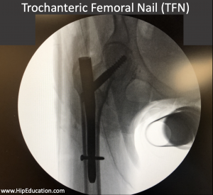
The trochanteric femoral nail procedure involves placing a metal rod into the bone through the greater trochanter, parallel to the shaft of the femur, to stabilize fractures of the proximal femur (figure 1). The trochanteric femoral nail is a specially designed metal rod with an associated hip screw.
Figure 1: X-ray of a trochanteric femoral nail
Aligning the fracture
The first step in the procedure is to realign the fractured ends of the bone (reduction) if they are not already aligned. This is essential for proper healing. This is done by placing the patient on a special operating table (fracture table) that allows traction to be applied to the injured leg. The leg is pulled and rotated and a special portable x-ray (fluoroscopy) is used to determine when the fracture has been reduced into an acceptable position.
Positioning the Trochanteric Femoral Nail
Then a K wire, or guide wire, is inserted from the outside of the hip (lateral approach) into the region of the greater trochanter and down the shaft of the thigh bone (femur) to help establish the positioning of the femoral nail. The canal in the middle of the thigh bone is then reamed to create enough space to hold the trochanteric femoral nail. The trochanteric femoral nail is hollow in the middle and this allows it to slide over the guide wire into the canal that has been created in the thigh bone. Once the nail is in acceptable position as determined by fluoroscopy the guide wire can be removed. The nail can either be “short”, extending just into the upper thigh, or “long” extending all the way down the length of the thigh bone (femoral shaft) to the knee. “Short trochanteric femoral nails” are used when the fracture is stable.
Positioning the Hip Screw
Once the nail has been placed into the femoral shaft, a screw, which enters through the outside of the femur is used to further stabilize the fracture. This “hip screw” runs through the trochanteric femoral nail into the center of the femoral head. A second screw is then interested into the far (distal) end of the trochanteric femoral nail to ensure that both ends of the nail are stabilized.
Recovery
Surgical procedures performed to treat intertrochanteric hip fractures usually require 3-6 months or longer to fully recover. This is longer than most other types of hip surgeries because the fracture area around the femur is the largest bone in the body and it therefore takes a relatively long time to form adequate new bone. Fortunately, the strength and stability of the trochanteric femoral nail usually allows the patient to bear full weight and undergo a focused rehabilitation program. Soon after surgery, walking weight bearing as tolerated using an assistive device such as crutches or a front-wheeled walker is encouraged to prevent stiffness and promote a full recovery. Range of motion exercises for the hip and knee should also be performed as soon as possible after surgery. The surgeon will prescribe physical therapy, which is designed to promote a return to pre-fracture activity levels and ensure that the full range of motion is regained.
Complications
Potential complications of trochanteric femoral nail placement include:
- Surgical site infection
- Post-operative abductor weakness and limp
- Blood clot: deep vein thrombosis and/or pulmonary embolism
- Failure of the bone fragments to rejoin (nonunion)
- Bone malrotation
- Malunion of the fracture

