Hip Anatomy
Bones and Joints of the Hip
The hip is a single ball-and-socket joint made up of two bones. These bones are the thigh bone (femur) and pelvis. The femur is a long bone that consists of a head, neck, shaft, and other protrusions such as the greater and lesser trochanter, which serve as attachment sites for muscles. One end of the femur contributes to the knee joint while the other contributes to the hip joint.
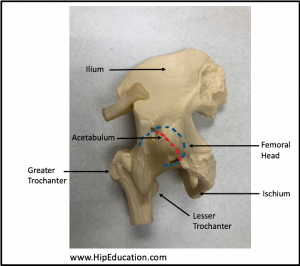
The pelvis is a complex structure shaped somewhat like a funny shaped bowl. It is formed from the fusion of 3 bones; the ilium, ischium, and pubis. One side of the hip joint is the head of the femur, which makes up the “ball”. On the other side is the “socket”, or acetabulum (figure 1). The acetabulum, which gets its name from a Greek word meaning vinegar cup, encircles the round head of the femur and creates a smooth surface over which the femur can glide. It is the meeting point of the 3 pelvic bones.
What is the purpose of the hip joint? The hip joint allows for standing, walking, and running. It allows for effective stabilization of the lower body, and transfer of force from the upper body to the lower body. The hip also allows us to move our upper legs in various ways in order to run, jump, and perform a variety of other activities.
Figure 1: Anterior view of the hip joint
Ligaments of the Hip
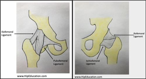
Ligaments are strong soft-tissue structures that span a joint connecting bones to other bones (figure 2). They provide stability and help keep the hip in place as the head of the femur moves in different directions within the acetabulum. The hip joint is stabilized by 5 ligaments:
- Iliofemoral Ligament (Y ligament of Bigelow)
- Ischiofemoral Ligament
- Pubofemoral Ligament
- Transverse Acetabular Ligament
- Ligamentum Teres (ligament of the head of the femur, foveal ligament, or round ligament of the femur)
Figure 2: anterior (left) and posterior (right) hip joint

The iliofemoral, ischiofemoral, and pubofemoral ligaments are named for the bones they connect: the ilium, ischium, and pubis. Together, they make up something called a joint capsule which surrounds and protects the hip. The transverse acetabular ligament connects the two ends of the lower acetabulum, and the ligamentum teres attaches the acetabulum to the head of the femur.
Muscles and Tendons of the Hip
Contractions of various muscles around the hip joint allow the thigh to move, or the pelvis to be stabilized. Muscles of the hip can be broken down into 6 categories based on the type of movement they allow the hip to make, Muscles can belong to more than one of these categories as they often allow for movement in more than one direction. In addition to movement, muscles of the hip also provide stability to the joint.
- Flexors – These muscles allow an individual to bend at the hip, as is done when bending to touch your toes. Hip flexors include the iliopsoas, rectus femoris, tensor fasciae latae, pectineus, adductor longus, and sartorius. They are generally located on the front or anterior side of the thigh.
- Extensors – Extensors allow the hip to “open” by shifting the leg behind the plane of the torso, like what happens to your back leg when trying to do a split. These muscles include the gluteus maximus, hamstrings muscle group consisting of the biceps femoris, semimembranosus, and semitendinosus, and the adductor magnus. Extensors are located on the back side of the thigh.
- Abductors – These muscles move the hip laterally, or out to the side of the body. This is consistent with their location on the outer side of the thigh. More commonly the abductor muscles serve to stabilize the hip when the other leg is off the ground. Muscles that abduct include the gluteus medius, gluteus minimus, tensor fasciae latae, piriformis, and sartorius.
- Adductors – These muscles move the upper legs inward (medially) toward each other. As such, they are located on the inner surface of the thigh close to the body’s midline and include the adductor longus, adductor brevis, adductor magnus, gracilis, and pectineus.
- External Rotators – External rotation refers to moving the hip out and away from the center of the body. Muscles that perform this action are at the back of the hip joint (posterolateral), or on the back of the thigh and far from the midline of the body. These include the gluteus maximus, piriformis, obturator internus, superior and inferior gemellus, and quadratus femoris.
- Internal Rotators – Internal rotation is movement of the hip inward toward the center of the body. Internal rotators are located posterolaterally, or on the back of the thigh and hip joint far from the body’s midline. These muscles include the gluteus medius, gluteus minimus, and tensor fasciae latae.
Each of the muscles discussed above is attached to bone via a tendon. Tendons are similar to ligaments in that they are made up of thick connective tissue. They are often sites of inflammation, or tendonitis, which is a major cause of hip pain
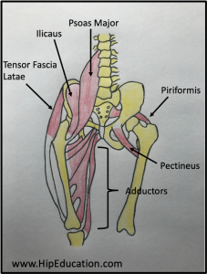
Figure 3: Drawing depicting the location of muscles supporting the hip joint.
Nerves of the Hip
There are many nerves that supply the hip, allowing us to perceive sensations such as pain and temperature, as well as signaling our muscles to move. Nerves originate from the spinal cord, traveling between spinal cord segments. After emerging from the spinal cord, they often fuse together and subsequently branch in order to supply various areas of the body. There are five main nerves whose branches supply the hip joint:
- Femoral nerve – Perceives sensation from the front, or anterior portion of the hip joint capsule
- Obturator nerve – Perceives sensation from the anterior portion of the hip joint capsule closer to the midline of the body, or anteromedially.
- Sciatic nerve – Perceives sensation from the back or posterior hip joint capsule, as well as the posteromedial area, or back of the hip closer to the midline.
- Nerve to quadratus femoris – Travels through the greater sciatic foramen and perceives sensation from the same areas as the sciatic nerve, the posterior and posteromedial hip joint capsule.
- Superior gluteal nerve – Travels through the greater sciatic foramen and continues on between the gluteus medius and minimus muscles. It detects sensory information from the posterolateral hip joint capsule, or back of the joint furthest away from the midline of the body.
Blood Supply of the Hip
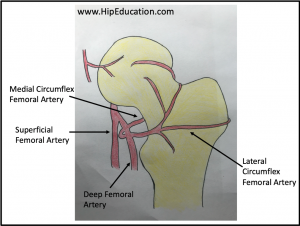
Adequate blood supply to the hip joint and its muscles is essential for proper hip function. The main blood supply to the hip comes from branches of the obturator and femoral arteries. The branches of the femoral artery that supply the hip include the following:
- Deep artery of the thigh – Supplies the adductor magnus muscle and the hamstrings muscle group
- Medial circumflex femoral artery – Supplies the head and neck of the femur, as well as the back of the thigh
- Lateral circumflex femoral artery – supplies the head of the femur and lateral thigh
The branches of the obturator artery that supply the hip include a posterior branch that delivers blood to the head of the femur and a branch that supplies muscles involved with adduction of the hip. Notably, there is a small branch of the obturator artery, the foveal artery, running through the ligamentum teres that can also supply blood to the head of the femur.
Figure 4: Blood supply to the femoral head


