
Hip Pinning
Indications
Hip pinning is used to treat femoral neck fractures. The femoral neck connects the shaft of the femur (thigh bone) to the ball of the femur (femoral head).Femoral neck fractures are commonly called intracapsular hip fractures because the femoral neck is located entirely within the capsule surrounding the hip joint.
Not all patients or all types of femoral neck fractures are good candidates for this surgery. Fractures that are well-aligned (non-displaced), impacted (stable and squished together), or that can first be effectively put back into place without surgery (closed reduction) are good candidates for hip pinning. In these situations, hip pinning holds the fracture in position until it heals. Displaced femoral neck fractures, especially in older patients, are usually treated with some sort of a hip replacement instead (partial hip replacement or total hip replacement).
Procedure
Hip pinning, also called percutaneous hip pinning, involves the placement of 3 screws through the upper part of the femur to hold a fracture together as it heals (figure 1). If the fracture is not aligned (displaced), the first step of the procedure requires lining up the two ends of the fracture so the bone can heal in its original location. This is called “closed reduction” and is typically performed under a special type of x-ray called fluoroscopy to better visualize the fracture.
Figure 1: X-ray showing the placement of hardware in a hip pinning
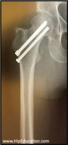
In most patients undergoing a hip pinning, the femoral neck fracture is stable and the surgeon can proceed directly to the second step which requires “fixing” the bone by holding it in place with screws. Before the screws are placed, wires called Krischner (K) wires are inserted through the two sides of the fracture to determine proper screw placement.
The correct position fo the wires and eventually the screws is determined by reviewing various fluoroscopic images of the hip taken at 90 degree angles to each other. Once the correct screw length and placement is determined using the wires, a cannulated drill (a drill with a hole through the middle) is used, and the wires are replaced by screws that travel perpendicularly to the fracture line and end up in the femoral head. These screws are tightened to hold the two ends of the fracture in place as it heals.
Recovery
The recovery period after hip pinning is generally longer than for other types of hip surgeries because it takes time for broken bone to heal. Typical recovery time is between 3-6 months. However, there is a large amount of variation in recovery time dependent on individual factors. The amount of weight that the patient can place through the affected leg after surgery will be determined by the surgeon. Often it is necessary to protect the healing femoral neck fracture by limiting the weight that the patient places on it. This will require the use of crutches or a walker for assistance. Regular range of motion exercises and physical therapy during the recovery period will help prevent stiffness and excessive muscle atrophy in the leg. Physical therapy also promotes a return to the level of activity that was possible before the fracture occurred.
Complications
Potential complications of hip pinning placement include:
- Avascular necrosis of the femoral head with resulting hip arthritis
- Failure of the ends of the fracture to heal (nonunion)
- Surgical site infection
- Prominence or protrusion of the screws.
Patient Reference Websites